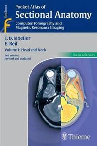
Pocket Atlas of Sectional Anatomy, Volume I: Head and Neck: Computed Tomography and Magnetic Resonance Imaging (Pocket)
avReif, Emil, Moeller, Torsten Bert, Meoller, Torsten B
ISBN: 9783131255044 - UTGIVEN: 2013-11-13This book is an authorized translation of the 3rd German edition published in 2011 by Georg Thieme Verlag, Stuttgart.[...]
Normal Findings in CT and MRI (Häftad)
avTorsten B. Moeller, Emil Reif
ISBN: 9780865778641 - UTGIVEN: 199909The key for any beginning radiologist who wishes to recognize pathological findings is to first acquire an ability to distinguish them from normal ones. This outstanding guide gives beginning radiologists the tools they need to systematically approach and recognize normal MR and CT images.Highlights[...]
Pocket Atlas of Sectional Anatomy, Volume 1: Head and Neck (Häftad)
avTorsten B. Moeller, Emil Reif
ISBN: 9781588904751 - UTGIVEN: 200610Now with all new images Known to radiologists around the world for its superior illustrations and practical features, the "Pocket Atlas of Sectional Anatomy" now reflects the very latest in state-of-the-art imaging technology. In the classroom and the clinic, this compact book acts as a highly spec[...]
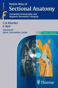
Pocket Atlas of Sectional Anatomy, Volume 3: Spine, Extremities, Joints (Häftad)
avTorsten B. Moeller, Emil Reif
ISBN: 9781588905666 - UTGIVEN: 200612Known to radiologists around the world for its superior illustrations and practical features, the "Pocket Atlas of Sectional Anatomy" now reflects the very latest in state-of-the-art imaging technology. In the classroom and the clinic, this compact book is a highly specialized navigational tool for [...]
Pocket Atlas of Sectional Anatomy, Volume II: Computed Tomography and Magnetic Resonance Imaging (Häftad)
avTorsten B. Moeller, Emil Reif
ISBN: 9781588905772 - UTGIVEN: 200611The second of a three-volume set which identifies the anatomical details visualized in the sectional imaging modalities. As a comprehensive reference, it is a great aid when interpreting images; schematic drawings of great clarity are juxtaposed with the CT and MRI images; anatomic structures are co[...]
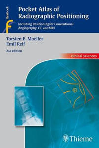
Pocket Atlas of Radiographic Positioning (Häftad)
avTorsten Moeller, Emil Reif
ISBN: 9783131074423 - UTGIVEN: 200811Praise for this book:Remarkable...a valuable, easy-to-use desk or pocket reference for medical imaging professionals at every level. - ADVANCE for Imaging & Radiation Oncology Now in its second edition, Pocket Atlas of Radiographic Positioning is a practical how-to guide that provides the detailed [...]
Taschenatlas der Schnittbildanatomie 2. Thorax, Abdomen, Becken (Häftad)
avTorsten Bert Möller, Emil Reif
ISBN: 9783131108036 - UTGIVEN: 2009-10Pocket Atlas of Sectional Anatomy (Häftad)
avTorsten Moeller, Emil Reif
ISBN: 9783131255037 - UTGIVEN: 200610Reflects the state-of-the-art imaging technology. In the classroom and the clinic, this compact book is a navigational tool for radiologists on the road to diagnostic success.[...]
Pocket Atlas of Sectional Anatomy (Häftad)
avTorsten Moeller, Emil Reif
ISBN: 9783131256034 - UTGIVEN: 2006-11Part of a three-volume set which identifies the anatomical details visualized in the sectional imaging modalities. As a comprehensive reference, this work is an aid when interpreting images; schematic drawings are juxtaposed with the CT and MRI images; anatomic structures are color-coded in the draw[...]
Pocket Atlas of Sectional Anatomy, Volume II: Thorax, Heart, Abdomen and Pelvis: Computed Tomography and Magnetic Resonance Imaging (Pocket)
avTorsten Bert Moeller, Emil Reif
ISBN: 9783131256041 - UTGIVEN: 2013-09-04This pocket atlas describes the anatomic details of sectional imaging in a concise, vivid format using radiology-specific terms, providing quick and easy access to vital information. The structures of the thorax, the abdomen, the pelvis and (in this new edition) the heart are illustrated by repre[...]
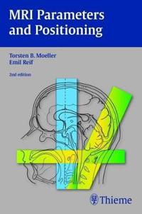
MRI Parameters and Positioning (Häftad)
avTorsten Moeller, Emil Reif
ISBN: 9783131305824 - UTGIVEN: 201002Packed with information on the practical aspects of MRI, this user-friendly text covers everything from advice on optimal positioning of patients to recommendations for setting the appropriate scanning parameters. Each consistently organized chapter follows the chronology of a standard procedure - [...]
Pocket Atlas of Sectional Anatomy (Häftad)
avTorsten Moeller, Emil Reif
ISBN: 9783131431714 - UTGIVEN: 200611Reflects the state-of-the-art imaging technology. In the classroom and the clinic, this compact book is a specialized navigational tool for radiologists on the road to diagnostic success.[...]
Atlas of Sectional Anatomy (Inbunden)
avTorsten B. Moeller, Emil Reif
ISBN: 9783131465412 - UTGIVEN: 2009-11-01A handy, full-color resource for interpreting musculoskeletal MRI scans with confidence This superbly illustrated atlas provides a comprehensive presentation of the normal sectional anatomy of the musculoskeletal system to aid in the diagnosis of diseases affecting the joints, soft tissues, bones, [...]
Taschenatlas der Röntgenanatomie (Häftad)
avTorsten Bert Möller, Emil Reif
ISBN: 9783137627043 - UTGIVEN: 201012Pocket Atlas of Radiographic Anatomy (Häftad)
avTorsten Moeller, Emil Reif
ISBN: 9783137842033 - UTGIVEN: 201004In this easily accessible pocket atlas, two expert radiologists present the normal radiographic anatomy readers need in order to interpret conventional diagnostic radiographs. Each practical, two-page unit displays a standard radiograph of a different projection on the left-hand side supplemented by[...]


